
Reabsorption
CSF absorption and drainage is essential in order to maintain the homeostasis of CSF pressure within the CNS. An imbalance between production and drainage can cause a significant rise in pressure resulting in reduced cerebral blood flow and therefore oxygen supply to the brain. At a molecular level increased pressure can denature proteins important for the function of neurons and glia. This emphasises the importance of the specialised epithelial cells which maintain cerebrospinal fluid volume. Regulation can be achieved by two mechanisms:
- The down-regulation of production by the choroid plexus.
- The up-regulation of absorption/drainage mechanisms.
An average human has between 150-160ml of CSF in their CNS at any one point, with approximately 500ml generated every day, so the mechanisms which maintain the volume within 10ml must be highly regulated. A reduction in the volume of CNS (which can be created artificially via a lumbar puncture) results in severe headaches, due to the increased effective net weight of the brain pushing on nerves and blood vessels at the base of the skull. This is turn can also lead to reduced cranial blood flow and eventually neuronal death.
The choroid plexus has ion and Aquaporin channels able to regulate the volume of CSF which they produce (see Fluid Secretion), so not only can they increase the volume of fluid, but also reduce it by limiting the rate at which CSF is produced. However greater contribution to reabsorption is made by the arachnoid villi.
Arachnoid Villi
The arachnoid villi (also known as arachnoid granulations) are protrusions of the arachnoid membrane which span into the dura mater. The dura mater contains many venous sinuses (which drain blood from the brain). This allows CSF to cross the arachnoid membrane and exit the subarachnoid space via the venous system. A major site of CSF reabsorption via arachnoid villi is the superior sagittal sinus (Blood vessels which span the top and back of the skull, draining deoxygenated blood from the brain). The mechanism of action for the villi is still not clear but it is thought that each granulation contains a one-way valve allowing the flow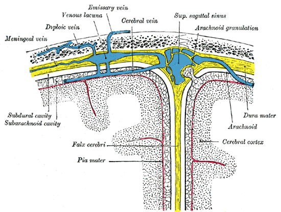 of fluid down a positive hydrostatic pressure gradient from the subarachnoid space into the low pressure venous system. Aquaporin channels have also been localised to the arachnoid villi and are thought to be essential for the movement of water across the blood-brain barrier. Current research hypothesises that arachnoid villi are important in reducing CSF volume and pressure when homeostasis is lost, but are not the main contributor to drainage under normal conditions. The main contributor is thought to be lymphatic drainage.
Dural Sinuses image courtesy of Wikimedia Commons under the creative commons license.
Lymphatic Drainage
Cerebrospinal fluid can drain directly into the lymphatic system via several routes including:
- Through the cribiform plate of the olfactory system.
- Along spinal nerves.
Cribiform plate
The Cribiform plate is part of the ethmoid bone containing many foramina through which the olfactory nerves descend into the nasal cavity. The CSF is able to pass through the cribiform plate and along the olfactory nerves into the cervical lymphatic system. There is evidence to suggest that this route is a major contributor to CSF drainage at normal intracranial pressure (obstruction of the cribiform plate severely impairs CSF reabsorption.
Spinal Nerves
Spinal nerves are present along the spinal cord and lie superficial to the central canal along its entire length. It is thought that CSF is drained from the canal via the spinal nerves. The CSF is passes over ruptured ependymal cells (see Production) and dorsal columns (sensory nerve pathway) then along the spinal nerves and into the dural lymphatic vessels of the spinal cord.
Hydrocephalus
A blockage of these reabsorption pathways can result in a condition known as hydrocephalus (increased volume of cerebrospinal fluid within the ventricles and subarachnoid space). Hydrocephalus can cause: increased intracranial pressure, enlargement of the head (in infants), convulsion, mental disability and eventually death. Infection or traumatic head injury can also cause hydrocephalus, as this too can affect the normal drainage of CSF.
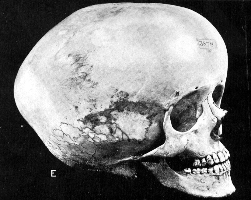
Hydrocephalic Skull image courtesy of Wikimedia Commons under the creative commons license.